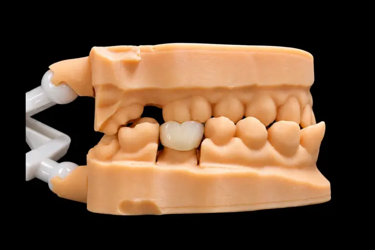
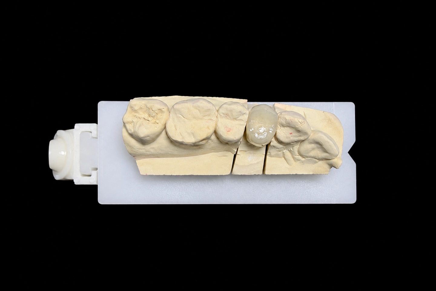
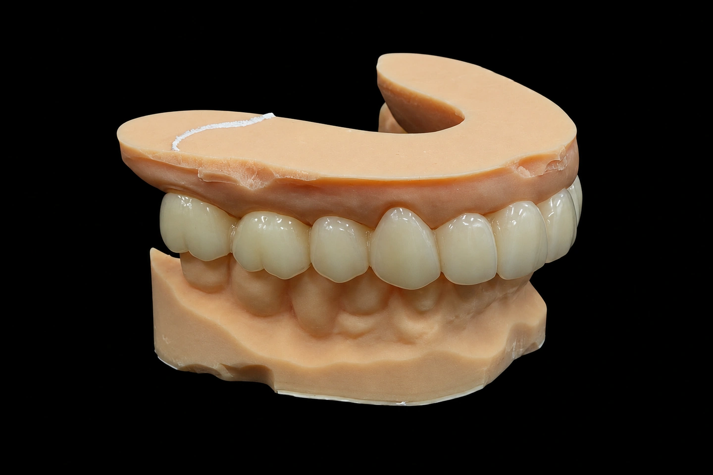
Patient situation
A 42-year-old male patient presented with a failed composite restoration on tooth #18 (mandibular left second molar). The composite had been placed 14 months earlier, at the same appointment as endodontic treatment. The restoration fractured and dislodged while eating.
The patient reported three specific complaints:
Clinical findings at the time of crown preparation:
The patient’s stated priority, recorded in the chart: “I want something that won’t break again — I don’t care what it looks like back there.”
The tooth had already been endodontically restored eight months prior; no post had been placed. The dentist prepared the tooth for a full-coverage crown with a 1.0 mm shoulder margin, equigingival on mesial and buccal walls, and subgingival 0.5 mm on the distal where the wall was compromised.
Occlusal clearance measured 1.2 mm at the central fossa in maximum intercuspation.
What the dentist ordered
The case arrived with a digital scan, bite registration, and a one-page prescription. No diagnostic wax-up, no provisional crown.
The dentist’s specifications:
Material
- PFM crown (porcelain-fused-to-metal)
- Cobalt-chromium framework, nickel-free
- Porcelain shade A3, to match adjacent tooth #17
Margin and preparation
- 1.0 mm shoulder on all walls
- Equigingival on mesial and buccal
- Subgingival 0.5 mm on distal (compromised wall)
- Adequate occlusal reduction for full-coverage crown
- Occlusal clearance: 1.2 mm, verified in centric occlusion
Restoration design as requested
- Porcelain on occlusal surface (this would become the critical constraint)
- Porcelain on buccal surface, shade A3
- Normal contact tightness at mesial and distal, with attention to preventing food impaction
- Balanced occlusal contacts in centric; no working-side interference
Clinical context provided
- Patient priority: durability over esthetics
- Opposing tooth shows wear from probable nocturnal clenching
- Tooth #18 not visible during normal smile or speech
- Previous restoration failed at 14 months
The dentist requested porcelain on the occlusal surface. That single specification — combined with the 1.2 mm clearance — would determine everything that followed.
Where the arithmetic failed
We do not open CAD on a PFM crown posterior until the thickness budget adds up. On this case, it did not.
A porcelain occlusal surface on a second molar under a worn opposing arch requires 1.5 mm minimum veneering porcelain thickness to survive functional loading. The metal framework underneath needs 0.4 mm. Cement space requires 0.05 mm.
The total requirement:
0.4 mm metal + 1.5 mm porcelain + 0.05 mm cement = 1.95 mm Available clearance: 1.2 mm Shortfall: 0.75 mm
A 0.7 mm porcelain occlusal layer does not fail gradually. It chips, and it chips early. This failure mode appears repeatedly in clinical case discussions — not because of material defects, but because of insufficient thickness.
The case could not be fabricated as prescribed. That had to be resolved before design began.
What the dentist decided
We contacted the dentist with the thickness calculation and explained the constraint: the available 1.2 mm would not support a porcelain occlusal surface on a molar under clenching load.
After reviewing the patient’s priority (durability) and the clinical context (posterior location, modest esthetic demand, history of restoration failure), the dentist made the following decisions:
Primary modification
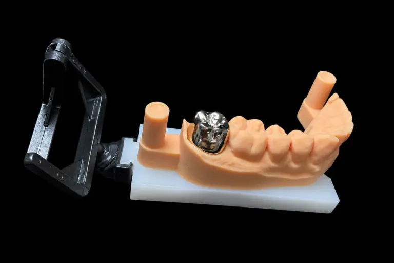
- Change occlusal surface from porcelain to metal
- Retain PFM construction (not switch to monolithic zirconia)
- Keep porcelain on buccal surface for acceptable appearance
Rationale documented in the dentist's response
- Tooth #18 is functionally critical but esthetically non-critical
- Patient explicitly prioritized “won’t break again” over appearance
- Metal occlusal surface eliminates chipping risk entirely
- Buccal porcelain maintains acceptable appearance in the limited visibility this tooth has
Additional specifications following the modification
- Metal occlusal surface to be polished (not glazed) for wear resistance
- Buccal porcelain shade A3 maintained
- Balanced centric contacts as originally requested
- No interference on lateral excursions
The dentist confirmed the plan with the patient before proceeding. The patient accepted the metal occlusal surface without concern.
How it was built
The fabrication followed the modified specifications, with each step aligned to the dentist’s requirements and the constraints imposed by 1.2 mm clearance.
Framework fabrication: We milled the cobalt-chromium framework from a disc blank using 5-axis wet milling, rather than casting. The reason: the occlusal needed to hold approximately 1.15 mm (to leave 0.05 mm for cement), while the buccal substructure had to stay under 0.4 mm to allow porcelain coverage. That dimensional control is reliable with milling; casting introduces variance we could not accept on a case with no thickness margin.
The framework was designed with:
After milling, the framework was verified with callipers at eight measurement points: five across the occlusal, three on the buccal wall. Margin integrity was checked under 10× magnification on the die. Any dimension outside tolerance would have returned the framework to the mill before porcelain application.
Surface preparation for porcelain bonding
- Aluminium oxide blasting at controlled pressure to create mechanical retention
- Ultrasonic cleaning in distilled water to remove residual abrasive and surface oils
- Pre-oxidation firing under vacuum to develop the oxide layer
The mechanical key from blasting provides primary retention; the oxide layer serves as secondary insurance against delamination.
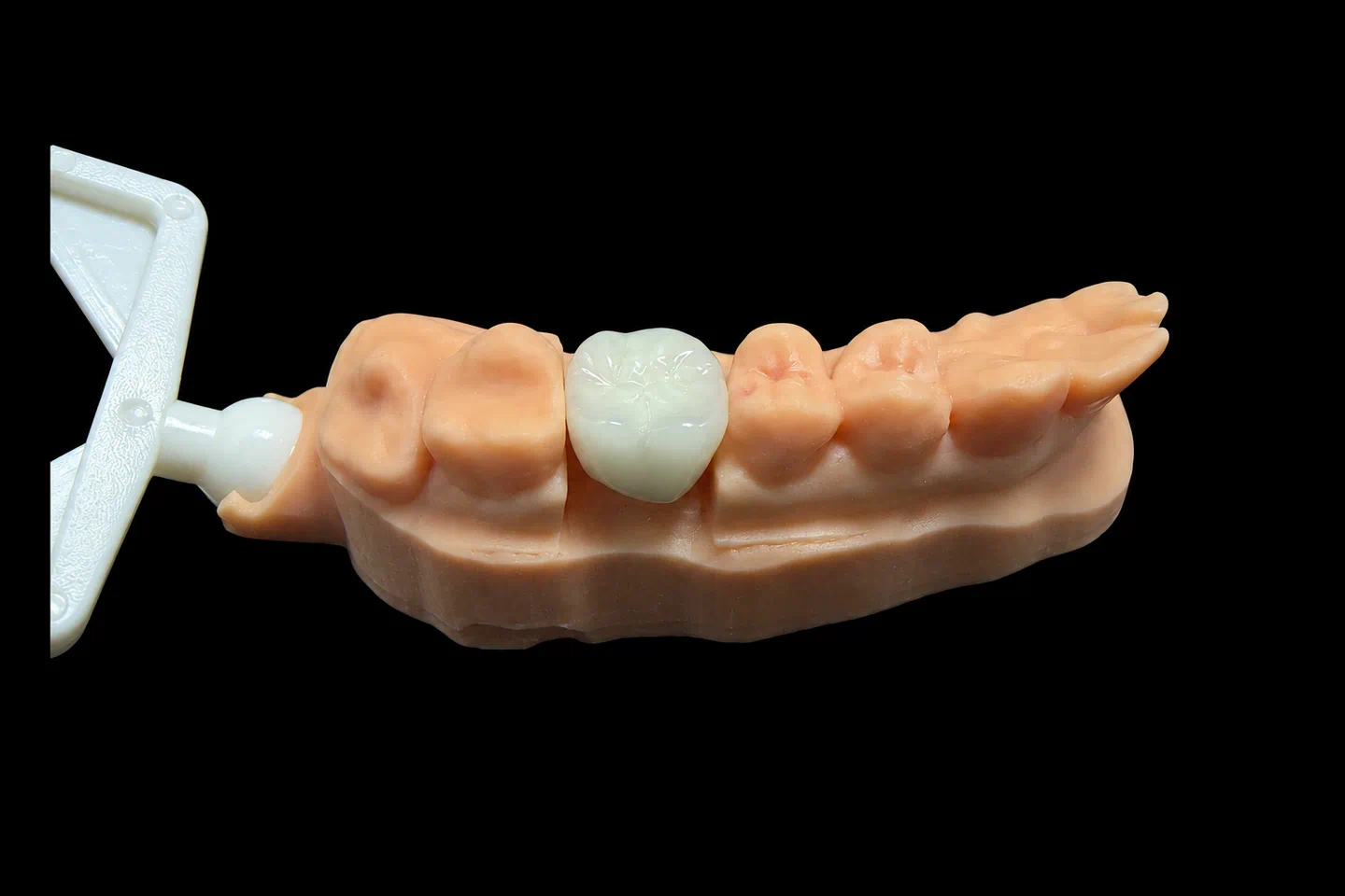
Porcelain application: Buccal surface only, shade A3 to match adjacent tooth #17. Three firing cycles:
No fourth corrective firing. Each cycle adds cumulative shrinkage and thermal stress to the thin framework. The body layer was applied slightly over-contoured to absorb expected sintering shrinkage, then contoured to final form before glaze firing rather than corrected afterward.
The buccal porcelain thickness measured approximately 1.0 mm after final firing — below our standard 1.5 mm target for posterior units, but sufficient for the buccal axial surface where it is not subject to occlusal loading.
Occlusal surface finishing: Metal only, as specified. Occlusal contacts were marked in centric occlusion and verified in lateral and protrusive excursions. One premature contact on the mesio-buccal cusp was identified and relieved.
The metal surface was finished progressively: coarse silicon carbide to establish final contour, medium and fine abrasives to remove machining marks, then diamond polishing paste to final mirror finish.
No glaze was applied to the occlusal. Polished cobalt-chromium offers superior wear resistance under bruxing load compared to glazed porcelain or even glazed metal.
Final verification before shipment
- Margins checked for complete seating on the die
- Occlusal contacts re-verified after glaze firing (sintering displaces porcelain; a crown that fits before glaze may not fit after)
- Proximal contacts checked for normal resistance
- Buccal contour inspected for smooth emergence from the margin
The completed crown was packaged with:
Turnaround: 3 working days from scan acceptance to shipment.
Delivery
The crown seated without adjustment at the delivery appointment.
The dentist confirmed:
The patient was informed that the occlusal surface was metal rather than porcelain, and the reason: to eliminate the chipping risk that had been calculated before fabrication. The patient expressed no concern — the stated priority had been durability.
The crown was cemented with resin-modified glass ionomer cement.
Patient response
The patient returned for a two-week follow-up appointment.
Functional outcomes
- Chewing comfort fully restored on the left side
- No food packing between #17 and #18
- No sensitivity to temperature or pressure
- No awareness of the restoration during chewing, speaking, or normal function
Patient's subjective assessment
The patient noted the metal occlusal surface and commented that it “looks industrial, but I don’t care — I can eat on it and it doesn’t hurt.” The priority established before fabrication (durability over appearance) was met.
Dentist's assessment
The dentist’s note to the lab after the follow-up visit: “Fits well, patient happy, no adjustments needed.”
If you are sending a case like this one
Three factors determine whether a PFM crown posterior is fabricable as prescribed:
1. Occlusal clearance
— measured, not estimated. Below 1.8 mm, a porcelain occlusal surface becomes a phone call. We will propose metal occlusal or material substitution, but we will not proceed silently and hope the porcelain holds.
2. Margin legibility in the digital scan
— especially for subgingival preparations. A subgingival shoulder or deep chamfer often produces a soft, ambiguous finish line in the scan. If we cannot trace the margin with confidence, we will ask for a re-scan or for guidance on where to place the margin. Guessing costs remakes.
3. Opposing dentition
— what the crown will occlude against. Natural enamel, composite, or metal is workable. A ceramic antagonist against a porcelain occlusal on a patient with evidence of bruxism or clenching is not — one surface will wear the other prematurely, and it is unpredictable which one loses.
Send those three details with the prescription and the case moves without interruption. Omit any of them and we will call, which delays both sides.