Why a palatal position is not a choice — it is forced anatomy
A maxillary premolar extraction socket is narrow, and the tooth’s long axis rarely aligns with the bone. A CBCT study of 179 patients and 716 maxillary premolars modelled two trajectories: implants placed along the original tooth axis (prosthetically driven) and implants placed where the bone best tolerates them (bone driven).
In the prosthetically driven position, 84.1% perforated cortical bone, 13.7% came within 2 mm of an adjacent structure, and only 2.2% — 16 of 716 — were clear of anatomy. Even optimised bone-driven placement still saw 40.5% perforation and 36.6% further compromise, leaving 22.9% genuinely uncomplicated.
The mean angle between those two trajectories — the implant-line A angle, ILAA — was 18.3° ± 8.0°, significantly larger for second premolars than first.
When we recovered this case and found the fixture positioned slightly palatal, we were not seeing a problem. We were seeing the outcome of a compromise that anatomy forces in the vast majority of cases at this site. The implant went where bone was available, not where the tooth used to live. Everything we subsequently had to solve in the laboratory was downstream of that decision.
The geometric ledger: five conditions that had to hold
For a screw-retained crown to work on a palatally inclined fixture, five things must happen simultaneously. We list them here because we state them before opening the design file — not after. The published literature does not address any of these, which is why this case matters to the laboratory.
1. The abutment axis must reach the occlusal table within tolerance.
A CBCT study of 58 implants in 47 patients — the only study that models this restoration type specifically in the premolar region — used a screw hole of 2.4 mm outer diameter. That creates an acceptable window of 1.2 mm from each cusp tip. Using this geometry, screw-and-cement restoration was feasible in 52% of first premolar sites and 78.8% of second premolar sites.
Our case fell into the 78.8%. We knew it was geometrically possible; we did not yet know where the margin of error lay.
2. The angle between the ridge axis and the abutment axis must stay below a threshold.
The same study quantified this as RA — the angle between the alveolar ridge’s long axis and the abutment axis. RA predicted the need for restoration type with 93.1% accuracy. Sites completed with screw-and-cement averaged 11.09° ± 3.69°; sites that required conventional cementation averaged 21.08° ± 5.04°. Each additional degree of RA raised the probability of needing cement retention by a factor of 1.885 (95% CI 1.31–2.70).
A palatal fixture inherently raises RA — it tilts the abutment axis buccally to catch the central fossa. The case was completed screw-retained, confirming it stayed within range. We did not measure RA independently, which means we cannot state the exact margin we held. That is a limitation we accept: without the clinician’s pre-operative scan file, we are working from surgical outcome, not surgical plan.
A structural note that matters for design strategy. The bone literature finds the second premolar harder: higher ILAA, higher perforation risk. The prosthetic literature finds it easier: 78.8% feasibility for screw-and-cement vs. 52%. No contradiction — they measure different problems. A palatal shift costs bone but buys occlusal table for the screw access. A laboratory that conflates surgical difficulty with prosthetic difficulty will either over-engineer easy cases or under-engineer hard ones. The two questions must be asked separately, and the answer to the second comes from a design file, not a radiograph.
3. The transmucosal height must be fixed before we design the contour.
The 1.75 mm collar went in at surgery and never moved. Soft tissue matured against a known, unchanging geometry for ten weeks. This is not a neutral detail — it is the foundation for the entire subsequent phase. If the provisional abutment had been exchanged or adjusted during healing, we would have had to predict contour from an anatomical socket, not copy it from tissue.
4. The emergence must be reproduced from the matured tissue, not extrapolated from the socket.
After ten weeks, the tissue has settled into a specific transmucosal form against the 1.75 mm collar. That form can be recorded. It cannot be reliably predicted from day-one radiographs. We took the impression on the provisional abutment rather than on the implant platform. The tissue did not have to collapse and re-form; it was captured in the state it had achieved.
5. Material thickness around the screw channel must be maintained as a specification.
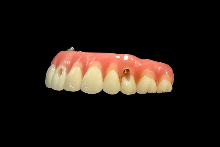
Once the screw access is offset to reach the occlusal table, ceramic thickness is no longer uniform. The distance between the channel wall and the external surface becomes an asymmetric constraint. We set a laboratory minimum for this dimension — there is no published consensus figure, and any laboratory quoting one is quoting a number it constructed. We record our minimum per case.
What we actually decided
The surgical team sent us a fixture positioned palatal, a 1.75 mm collar that would not be exchanged, and a request for screw retention. That request triggered four laboratory decisions, each with alternatives we considered and rejected.
Decision 1: Screw-and-cement retention vs. an angulated channel on a custom abutment
Two routes existed.
Route A: Design a custom abutment with an angulated screw channel. This shifts the access path; the crown’s emergence is still engineered in ceramic against the custom abutment body. This approach works well for modest divergence, provided the implant system permits it. The limit is system-specific correction range — and the cardinal error is using channel angulation to mask a severely malpositioned implant.
Route B: Cement the crown to a low-profile titanium base outside the mouth, then screw that assembly to the implant.
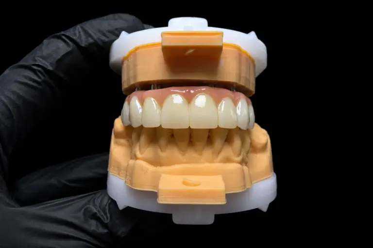
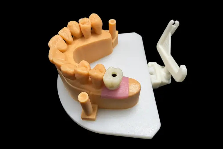
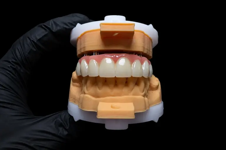
We chose B. Here is the structural logic: in Route A, one interface — between the abutment and the fixture — is left exposed in the mouth and must be cleaned up at the chair. In Route B, that interface stays protected inside the assembly, and the only junction the clinician sees is a screw. The only interface that cannot be inspected after delivery is the cement line between the crown’s intaglio and the titanium base — and that interface is entirely under our control, made outside the mouth, under direct vision, with any excess removable before seating.
At a second premolar site where screw-and-cement feasibility runs at 78.8%, this is the lower-variance path. It is not universal — section 6 states where it stops working — but for this case, it was the right call.
Decision 2: The provisional collar never moved. Tissue matured against it for ten weeks.
The surgical team placed a 1.75 mm collar at immediate loading and did not exchange it. We took that as a fixed constraint. If we had asked for the collar to be removed and reimplanted, we would have triggered tissue collapse and re-formation, and we would be predicting emergence geometry from radiographs and bone landmarks, not from tissue.
Instead, we left it untouched for ten weeks. At the re-entry appointment, we took the impression on the collar, not on the implant platform. This one decision eliminated the largest variable in soft-tissue transferability.
Decision 3: The emergence was recorded from the matured tissue, not designed from the socket
The conventional approach to premolar emergence is to begin at the clinician’s provisional or, if one is not available, to build emergence from surgical geometry — bone position, implant trajectory, imagined soft tissue.
We had a ten-week record of actual soft tissue form. We copied it. That is a different design posture entirely. It removes the most common emergence failure mode: over-building the subgingival bulge based on an assumption of what the tissue “should” be.
A systematic review of surgical and implant factors notes an observational association between an emergence angle ≥45° and peri-implantitis. This is a risk signal from observational data, not proof that one angle causes disease, and it does not specify a target. It does mean “bulk out the emergence until it looks full” is indefensible. We designed for a smooth transition rather than convex bulk, and we had tissue maturity on our side to verify it.
Decision 4: The marginal interface is now our responsibility, not the chair's
In this design, the cement sits between the crown’s intaglio surface and the titanium base collar. That interface is closed outside the mouth and stays closed. No cement ends up subgingivally in the patient’s mouth — the clinician’s only action is to seat and secure a screw.
The trade-off: marginal fit of that intaglio becomes entirely a laboratory variable. There is no clinician at the chair to adjust a marginal overhang, no radiographic check that catches excess cement. The fit has to be right before the case leaves our building. That is the literal sense in which screw-and-cement restoration is a laboratory restoration — it is built in our environment, under our specifications, and it arrives clinically ready.
What the one-year result tells us — and what it does not
At one year, the clinician reported soft tissue gain in the interproximal area and stable marginal bone.
That is the correct attribution and also the limit of what this case can teach. In play are flapless extraction with preserved bone walls, buccal grafting, immediate loading, palatal positioning, an untouched 1.75 mm collar, and tissue-recorded emergence. A single case cannot isolate these variables. Claiming the interproximal gain as proof of our contour design is a causal claim the case does not support.
What can be said is narrower and more useful: the sequence produced an outcome in which the laboratory’s contribution was not limiting. We did not cause the interproximal gain, but we did not prevent it. That is the honest reading, and it is actionable for a clinician.
Where this approach stops working: the boundary conditions
We hold three categories of limits: geometric limits, tissue-recovery limits, and patient-factor limits.
It worked here because
- Second premolar position — screw-and-cement feasibility is 78.8% at this site, vs. 52% at the first premolar. Geometry permitted the approach.
- Apical anchorage was adequate — minimum implant length requirements in the premolar socket range 6.5–19.0 mm; we had 13 mm in a socket that required placement. Primary stability was 56 Ncm at surgery.
- The provisional collar remained untouched for 10 weeks — soft tissue matured against a known geometry. This is non-negotiable for tissue-recorded emergence.
- Patient factors were favorable — good general health, smoker but without severe parafunction reported. These affect healing and long-term stability.
It would not have worked if
- The RA had climbed toward 20°+ — in the cement-retention group, RA averaged 21.08°. If our implant trajectory had required that much compensation, the screw channel would have migrated out of the 1.2 mm window, and we would have been forced to recommend conventional cementation pre-operatively.
- The first premolar had been the site — 52% feasibility is a coin toss on geometry alone. First premolar cases need individual analysis and conservative planning.
- The provisional had been exchanged or adjusted during healing — tissue contour then becomes an inference rather than a record, and all the gains of tissue-copy emergence vanish.
- Parafunction or severe occlusal overload had been reported — that changes the ceramic thickness requirement, the contact scheme, the entire stress model. Screw-and-cement retention does not inherently solve a bruxism case; it requires coordinated occlusal strategy.
One further boundary belongs to the case itself rather than the method: one year is an outcome, not a survival rate. Screw-and-cement restoration carries its own maintenance profile — screw loosening, cement retention loss, ceramic fracture at the junction. A one-year follow-up cannot speak to long-term incidence of these events.
What we still ask for before we design
This list is the practical output of our design logic. We ask for these five items with every premolar implant case because missing any one of them turns the case into a guess.
Send these five items plus a final scan and occlusal records, and the design conversation takes one exchange. Send only a scan and a shade, and it takes four.
Questions we receive about this case
Can a palatally positioned premolar implant still be restored screw-retained?
At the second premolar, usually — modelling shows feasibility in 78.8% of cases. At the first premolar, roughly half the time. The determinant is the angle between the ridge axis and the abutment axis (RA), not implant brand or diameter.
Why cement the crown to the abutment outside the mouth instead of ceasing it in the patient?
Because it eliminates the principal limitation of cement-retained restorations: inaccessible excess cement. The interface is completed under direct vision, excess is removed before seating, and the restoration remains retrievable with a screwdriver. The trade-off is that marginal fit becomes entirely a laboratory responsibility, with no chairside correction available.
What is the angular correction limit before the case must be cemented in situ?
There is no universal figure. Any laboratory quoting a single number across all systems is quoting a number it invented or borrowed from a single system. The usable constraint is geometric: a 2.4 mm screw hole must land within 1.2 mm of both cusp tips. Work that constraint backwards from the patient’s occlusal anatomy, and the available correction follows.