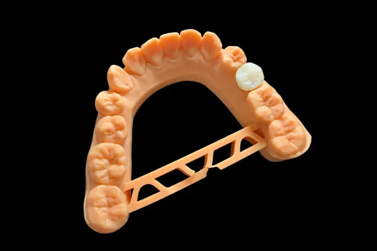
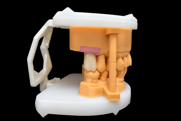
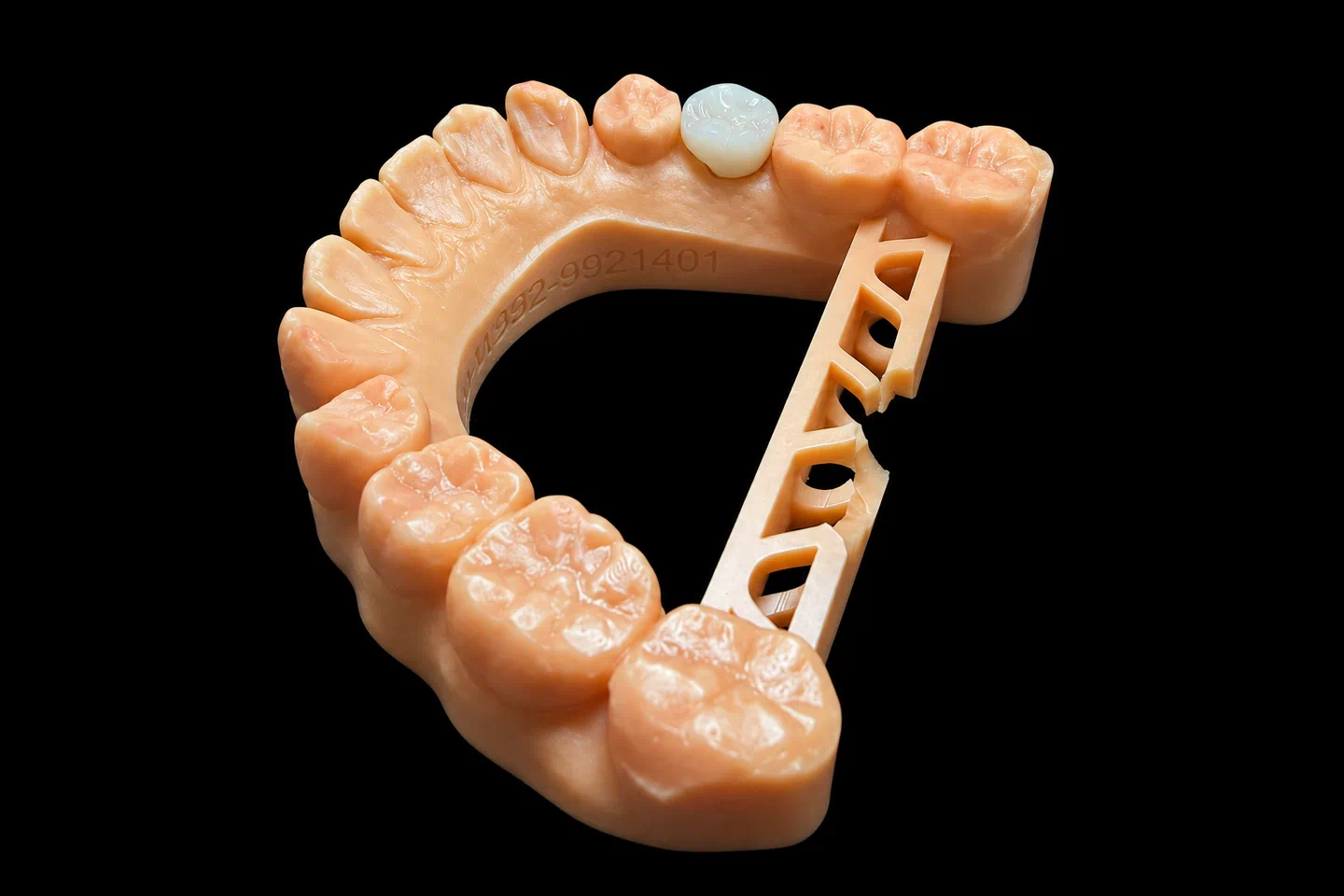
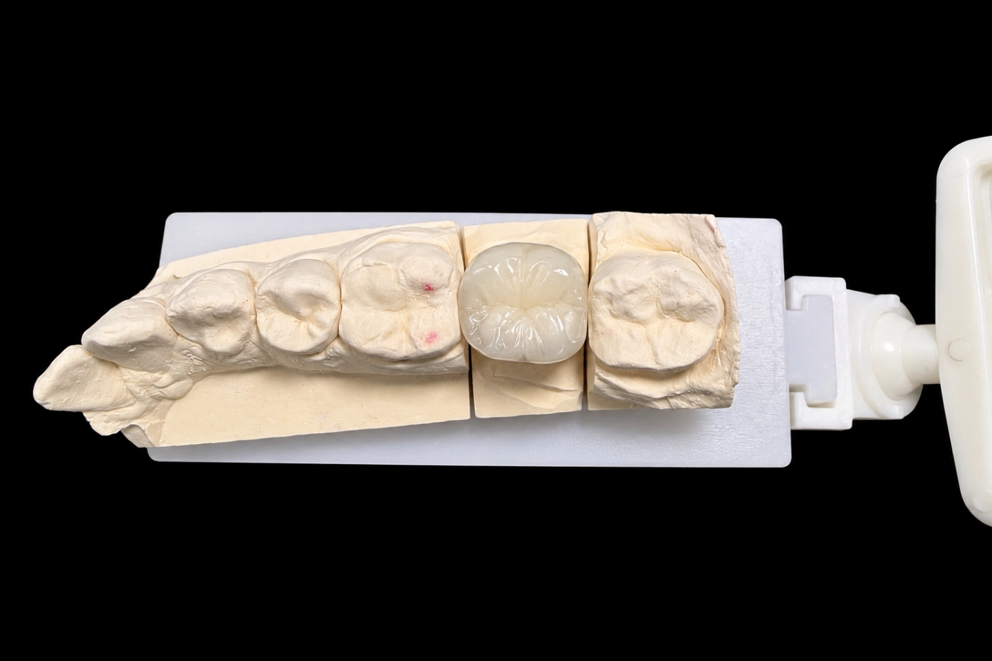
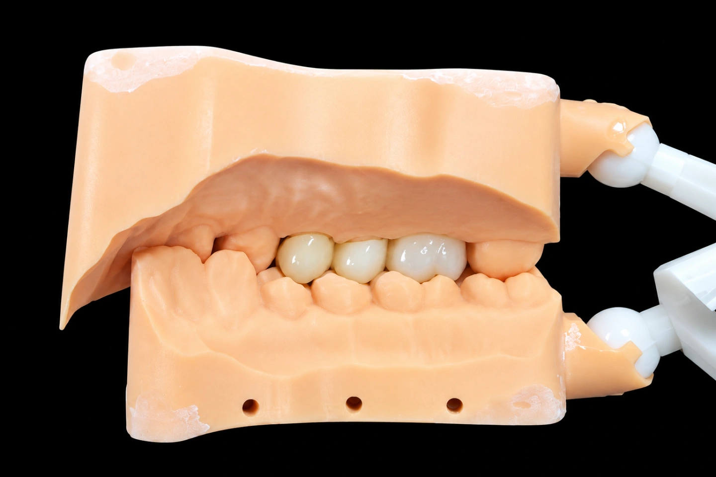
Patient Situation
The patient is 24 years old and was born without #10. Braces opened the space, and the orthodontist handed the case to the restoring dentist right after debonding . The dentist and patient chose a bonded anterior zirconia bridge over an implant. The root gap between #9 and #11 was narrow on the CBCT, and the patient didn’t want surgery .
What the dentist sent: intraoral scans of both arches in STL, a bite scan, scans of left and right side moves and of the front teeth sliding forward, the Rx, and shade photos of #9 with tabs held next to it .
What the Dentist Asked For
The Rx, condensed :
Requests 1 and 6 can’t both work on #11.
The Technical Problem: No Room Behind the Canine
What we measured
Our CAD technician set the bite scan and the side-move scans on the models. Then we measured the gap between the palatal surface of each abutment and the lower teeth, where a wing would have to sit .
| Where | Free room in the bite | Free room in side moves |
|---|---|---|
| #9, cervical half of the palatal face | 1.2 mm | Not touched in any move |
| #9, incisal third | 0 mm, lower incisors touch here | Touched in the forward slide |
| #11, cingulum area | 0.4 mm | 0 mm, on the guidance path |
A zirconia wing needs real thickness to be strong enough to bond and to handle the bite. On #11, a wing of usable thickness would stand right in the path of the lower canine. Then the canine would hit the wing first, and the left side move would change.
Why two wings carry a hidden risk
Two different teeth don’t move the same way under load. A central and a canine move a little differently when the patient bites, and a two-wing bridge ties them together. That puts stress on the bond line or on a connector.
When one wing lets go and the other holds, the bridge can still feel solid to the patient. Nobody notices the loose side until there’s a problem under it. With a single wing, a debond shows up right away: the bridge comes out, so the dentist knows the same day.
The clinical data points the same way. In a prospective study of anterior bonded bridges made from glass-infiltrated alumina, four of the 16 two-wing bridges broke at one connector within 15 months. The five-year survival was 73.9% for two wings and 92.3% for single wings. That was an older ceramic, not zirconia, but the pattern is why we raised the design question.
The Options We Sent Before Design Approval
We sent the dentist the measurements, a draft design and three options :
| Option | What it means | What you give up |
|---|---|---|
| A. Two wings, as written | Wings on #9 and #11 | The #11 wing changes the canine guidance, unless #11 is prepped deeper or the guidance is reshaped. Both are clinical calls. It also keeps the two-tooth link |
| B. Single wing on #11 | Cantilever off the canine | Same room problem as A, and the wing sits on the tooth that does the guiding |
| C. Single wing on #9 | Cantilever off the central | The wing has to stay below the lower incisor contacts, so its outline is smaller |
We recommended C. The dentist agreed and approved it by email . Then they prepped #9 to the wing outline we marked on the design preview, all in enamel: a light finish line near the gum, a small box on the distal side facing #10, and a shallow dimple in the middle of the palatal face . They sent the prep scan the next day .
How We Built the Anterior Zirconia Bridge
Prep scan check
Before final design, we put the prep scan back in the bite. We checked three things :
All three passed , so we moved on to design.
Design
Milling, sintering and fit check
We milled the bridge in one piece from a multilayer zirconia disc . The software scales the design up to make up for shrinkage in the furnace. Sintering runs on a fixed, logged program, and the bridge sat on a support tray so the cantilever couldn’t sag.
After sintering, the bridge went back on the printed model of the prep. We checked that the wing seated fully with no rocking, and that the edges met the finish line under magnification. Then came stain and glaze on the facial side, and step-by-step polishing on the palatal side.
Finished Result
The anterior zirconia bridge at final inspection :
| Check | Result |
|---|---|
| Wing thickness and connector size | Measured, matching the approved design |
| Seat on the model | Full, one path in, no rocking |
| Wing edges | On the finish line, no gaps under magnification |
| Pontic contacts on the articulated scans | None found |
| Shade | Checked against the #9 photos under standardized light |
| Turnaround | Shipped on working day 6 after the prep scan |
Feedback after the try-in
A summary of what the dentist told us after the try-in and the two-week check. The patient’s comments came to us through the dentist .
- The wing seated fully at the try-in with no adjustments, and the shade matched #9 with try-in paste. They bonded it the same visit under rubber dam with an MDP-containing resin cement.
- At two weeks the bridge was firm. The left side move still ran on #11 alone, and articulating paper left no marks on the pontic.
- Having the room measured before the prep meant no guidance changes at the chair.
- They liked how the new tooth sits next to #9, with no gray showing through.
- The wing behind #9 felt new for a few days, then stopped being noticeable. Speech felt normal from the start.
- Cleaning under the pontic with a floss threader took some practice at first, then became routine.
What Long-Term Data Says About Single-Wing Zirconia Bridges
Single-wing designs are the best-documented kind of anterior zirconia bridge:
These are pooled results from selected patients, not a promise for any one bridge.
Zirconia Bridge Front Teeth Designs We Make
For missing front teeth, we make three zirconia bridge designs:
If your Rx names a design that we see a risk with, we’ll tell you why and offer options. You decide. We never switch the design or the material without your OK, written or verbal.
Sending Us an Anterior Zirconia Bridge
Every step, from your data to QC and packing, happens in our own labs: four of them in Shenzhen and Huizhou, China, with about 460 people. Istar is an FDA-registered establishment, Registration Number 3039441248, which you can check in FDA’s establishment database. The lab runs under ISO 13485.
Have a missing incisor and you’re not sure which tooth should carry the wing? Send us the scans with the bite and side moves, and we’ll measure the room before anything gets made. Send your first case or talk to us.
FAQ
What shade photos help on a front-tooth bridge?
Take one photo with the neighboring natural tooth and shade tabs in the same frame. If you plan to prep, add one of the prepped tooth with a tab too. Camera settings, flash and filters all shift color, so keep them the same for every shot of the case.
Can you make a bonded anterior zirconia bridge for a patient who grinds?
Yes. We’ll flag the risk with the design and suggest options. If you still want it as written, we make it to your Rx. Problems caused by that design, including remakes, are then on your side.
Can I send my own CAD design for milling?
Yes. If you ask us to mill your design as-is, we mill it as sent, so please double-check it first. If we spot a clear problem, like a thin wing or connector, we’ll tell you before milling. If you still want it as designed, we make it to your file.
Do you take rush cases?
Yes, at no extra charge, for clients who send 10 or more cases a month. We guarantee the ship date. The arrival date depends on customs, so we can’t guarantee that.